Bones of the foot and ankle, medial view with labels App… Flickr
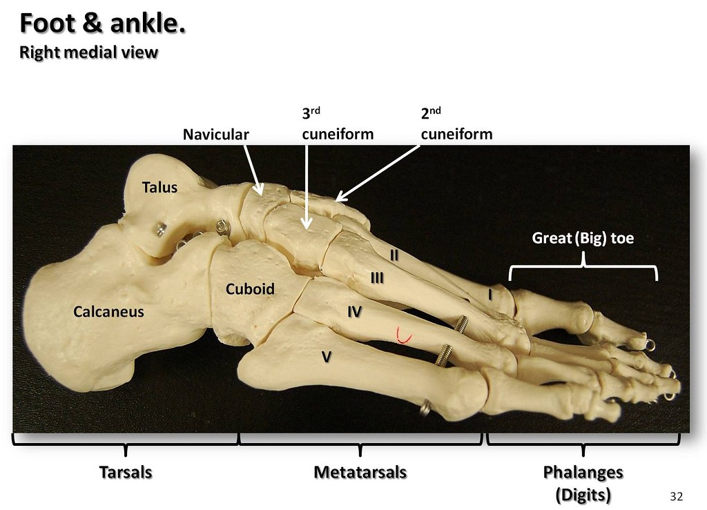
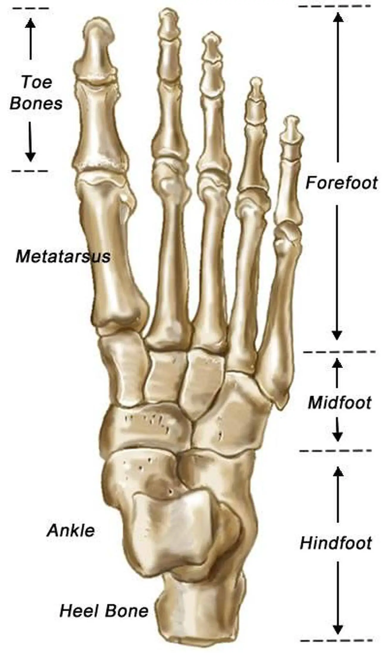
Introduction. The skeleton of the foot consists of 26 bones and these can be grouped into three groups:. The tarsus (ankle joint); The metatarsus; The phalanges (bones of the toes); There are 7 tarsal bones, 5 metatarsal bones and 14 phalanges.. Anatomically the foot can be divided into the forefoot (metatarsals and phalanges), the midfoot (cuboid, navicular and cuneiforms) and the hindfoot.

Foot bones anatomy Royalty Free Vector Image VectorStock
How many bones are in the foot? There are 26 bones in the foot and 33 joints in the foot. The foot is split anatomically into 3 sections; the hindfoot, the midfoot, and the forefoot. This article will describe in detail the anatomy and function of the major bones in the foot. Foot Bones: Hindfoot

Ankle Bones Diagram koibana.info Foot anatomy, Anatomy bones, Ankle
Fore-foot - the fore-foot is composed of the metatarsals and phalanges. The bones that comprise the fore-foot are those that are last to leave the ground during walking. Mobile Joints of the foot and ankle: (See Figure 3.) Ankle joint. Sub-talar joint. Talo-navicular joint. Metatarso-phalangeal (MTP) joints.

Pin on Anatomny
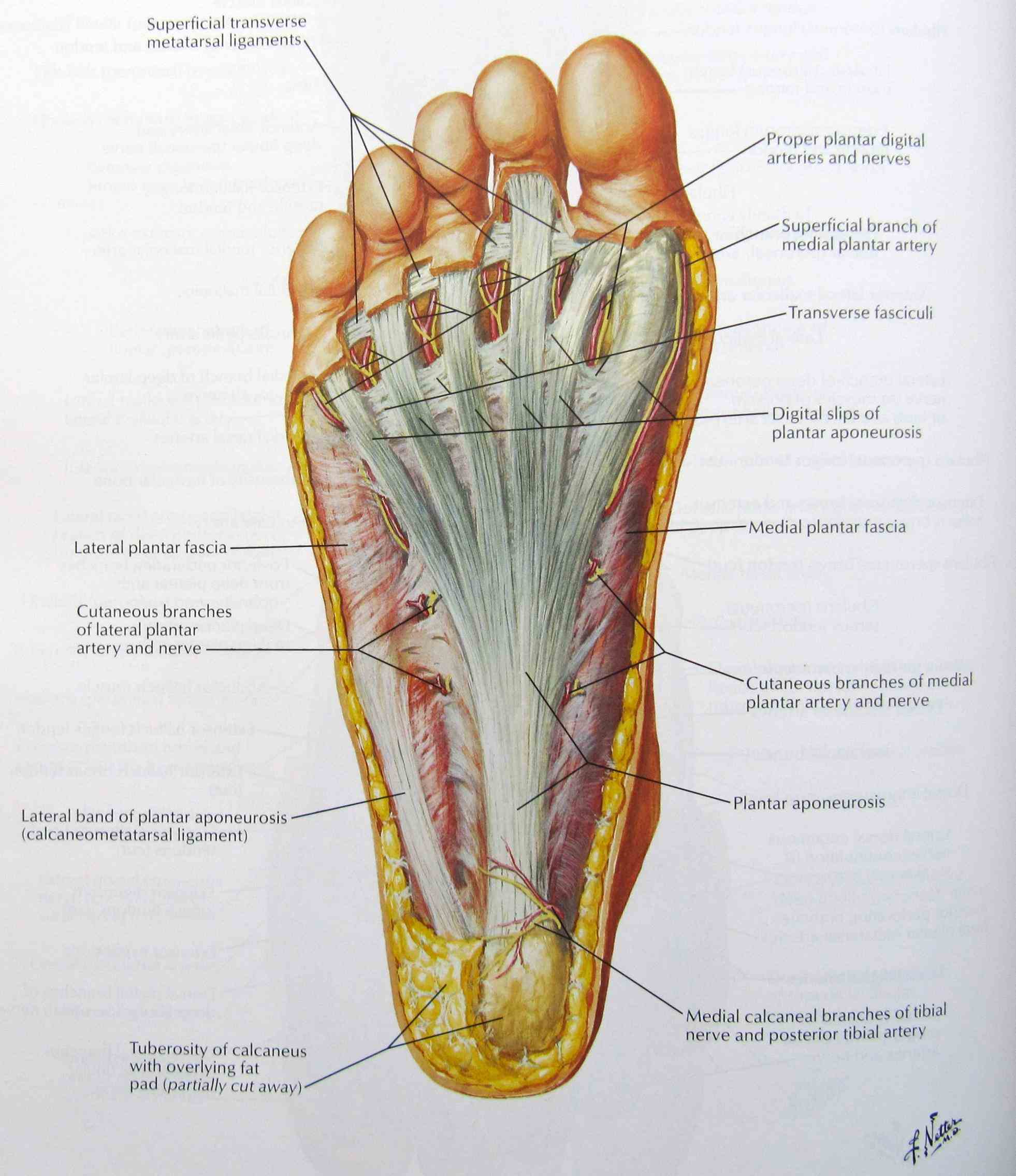
The foot is the region of the body distal to the leg and consists of 28 bones. These bones are arranged into longitudinal and transverse arches with the support of various muscles and ligaments. There are three arches in the foot, which are referred to as: Medial longitudinal arch. Lateral longitudinal arch.

The Cuboid is a cubeshaped bone present on the outer side of the
Foot Foot The foot is the lowermost point of the human leg. The foot's shape, along with the body's natural balance-keeping systems, make humans capable of not only walking, but also.

Foot treatment Orthopaedic Adam Budgen
Summary. A Jones fracture of the foot is a break in the fifth metatarsal bone. Symptoms may include bruising, swelling, pain, and difficulty or inability to walk. Treatment may include immobilizing the foot with a cast or medical boot or surgery to repair the break.

Pin by Susan Garverick on Education Medical anatomy, Anatomy bones
Cuboid Medial cuneiform Intermediate cuneiform Lateral cuneiform Some people may be born with an extra navicular bone ( accessory navicular) beside the regular navicular bone, on the inside of the foot. This is a normal anatomical variation seen in around 2.5% of the entire population of the US. Metatarsal Bones

anatomy of the foot Ballet News Straight from the stage bringing
Diagnosis Treatment The parts of the foot and its functions are unique but can also contribute to common foot problems. The many bones, ligaments, and tendons of the foot help you move, but they can also be injured and limit your mobility.

Foot Bone Anatomy Vector Illustration 539973 Vector Art at Vecteezy
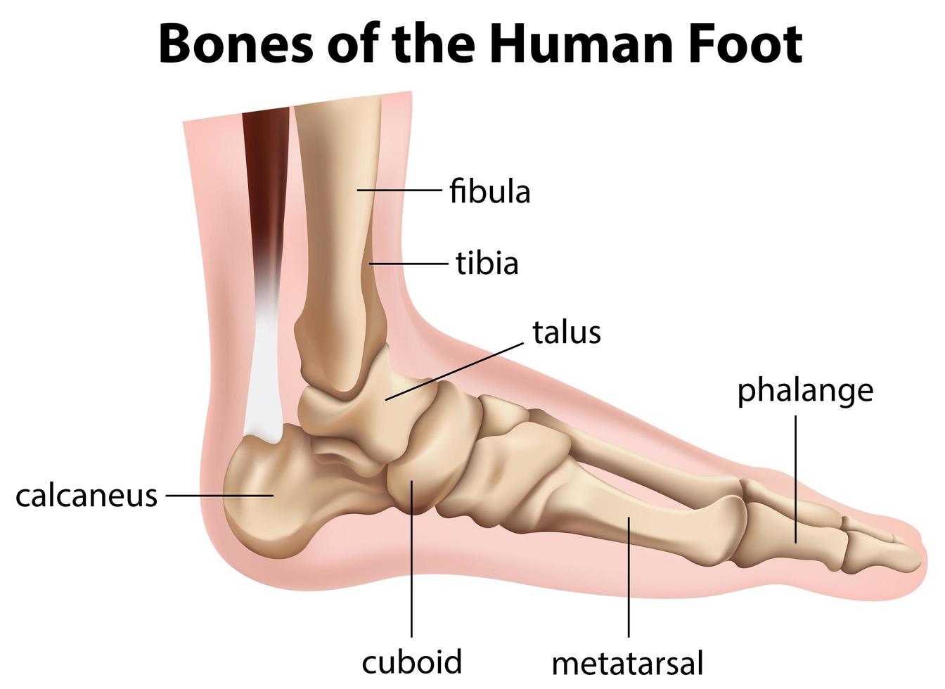
The diagram of bones in the ankle and foot is given below: Tarsal Bones The tarsal bones in the foot are located amongst tibia, metatarsal bones, and fibula. There are in all 7 bones, which fall under tarsal bones category. They are: Calcaneus or Calcaneum: To explain the term in layman's language, it is the heel bone in the skeletal system.
Anatomy The Bones Of The Foot
There are 26 bones in the foot, divided into three groups: Seven tarsal bones Five metatarsal bones Fourteen phalanges Tarsals make up a strong weight bearing platform. They are homologous to the carpals in the wrist and are divided into three groups: proximal, intermediate, and distal.
Bones of the human foot diagram 1142236 Vector Art at Vecteezy
The foot bones account for a quarter of all the bones in our body. Find out how the different foot bones fit together and how they are commonly injured. Home Diagnosis Diagnosis Guide Diagnosis Chart Top Of Foot Pain Ball Of Foot Pain Inner Foot Pain Outer Foot Pain Foot Arch Pain Heel Pain Toe Pain Nerve Pain Symptoms Symptoms Guide Blisters
Pictures Of Bones Of The Feet
Last updated 2 Nov 2018 The anatomy of the foot The foot contains a lot of moving parts - 26 bones, 33 joints and over 100 ligaments. The foot is divided into three sections - the forefoot, the midfoot and the hindfoot. The forefoot

Pin on Anatomy and physiology diagrams
Foot and ankle anatomy consists of 33 bones, 26 joints and over a hundred muscles, ligaments and tendons. This complex network of structures fit and work together to bear weight, allow movement and provide a stable base for us to stand and move on. The foot needs to be strong and stable to support us, yet flexible to allow all sorts of complex.

Foot & Ankle Bones
Columns of the Foot The foot is sometimes described as having two columns (Figure 3). The medial column is more mobile and consists of the talus, navicular, medial cuneiform, 1st metatarsal, and great toe. The lateral column is stiffer and includes the calcaneus, cuboid, and the 4th and 5th metatarsals. Figure 3: Columns of the Foot

Ankle and Foot Pain Massage Therapy Connections
The seven tarsal bones are: Calcaneus: The largest bone of the foot, it is commonly referred to as the heel of the foot. It points upward, while the remaining bones of the feet point.

The bones in the foot inferior view (Picture illustrated from Thieme
Use these bones of the foot quizzes to master your identification skills. Overview of the bones of the foot and their divisions into the hindfoot, midfoot and forefoot. With a total of 26 bones in each foot, learning the bony anatomy of the foot is no piece of cake. That is, the memorization aspect.